Interactive VR Assists in Treatment PlanningBy HospiMedica International staff writers[16 Apr 2018] 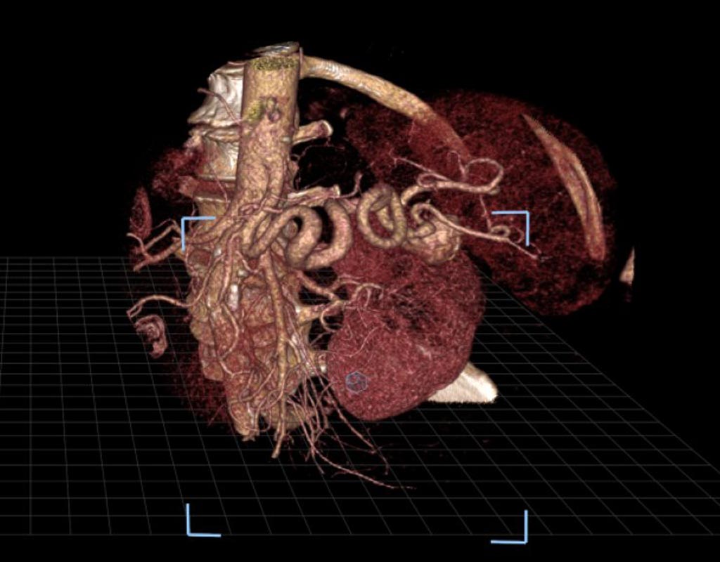 |
|
| Interactive virtual reality (VR) shows a patient's unique internal anatomy to interventional radiologists in order to help physicians effectively prepare and tailor their approach to complex treatments, such as splenic artery aneurysm repair. In a new study, researchers compared the VR technology to the use of images from a commonly used visualization software system that displays images on a standard two-dimensional platform and found the accuracy to be similar with both methods, though confidence improved substantially with VR. VR converts a patient's pre-procedural CT scans into 3D images that can be virtually moved and examined by radiologists wearing virtual reality-type glasses. VR allows operators to manipulate routine, two-dimensional images in an open three-dimensional space in order to allow them to look into the patients' organs and tissues that had been impossible outside of the human body, until now. This arms the operators with a deeper and intuitive understanding of spatial relationships, such as between an aneurysm and the surrounding arteries. In the study, three radiologists used both the technologies to independently evaluate 17 splenic artery aneurysms in 14 patients. The researchers measured the accuracy of the radiologists in identifying inflow and outflow arteries associated with the aneurysms with each method. The radiologists also ranked improvements in their confidence on a four-point scale when using VR as compared to the standard method. The researchers found that 93% of the participating physicians who used the VR method indicated higher confidence in their abilities (a score of at least three). "Treating splenic artery aneurysms can be very difficult because of their intricate nature and anatomic variations from patient to patient. This new platform allows you to view a patient's arterial anatomy in a three-dimensional image, as if it is right in front of you, which may help interventional radiologists more quickly and thoroughly plan for the equipment and tools they'll need for a successful outcome," said Zlatko Devcic, M.D., a fellow of interventional radiology at Stanford University School of Medicine and collaborating author of the study. "Pre-operative planning is possibly the most important step towards successfully treating a patient, so the value of VR cannot be understated," Devcic said. "This technology gives us a totally different way to look at that structure and safely plan our approach to patient care." The research was presented at the Society of Interventional Radiology's 2018 Annual Scientific Meeting. The researchers hope that more studies in the future will examine whether the VR technology will ultimately help reduce the time required to perform the treatment and consequently, reduce the amount of radiation and contrast exposure to the patient. |
|